
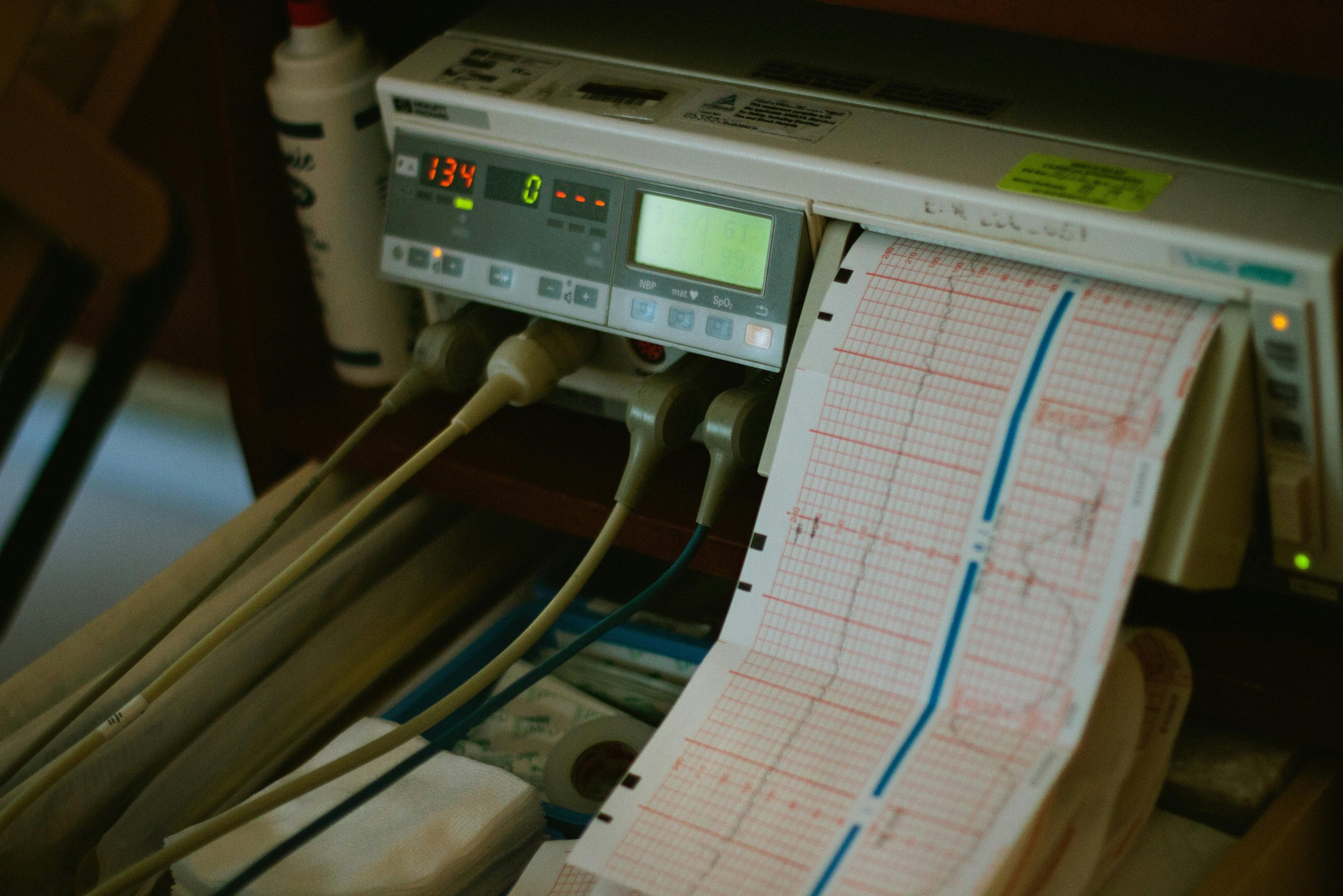
Diagnostic Testing
Our cardiac diagnostic testing helps assess, monitor, and support the management of heart conditions. We aim to provide accurate, timely results in a supportive environment, helping you and your doctor make informed decisions about your care.
-
An echocardiogram (often called an “echo”) is a non-invasive test that uses ultrasound (high-frequency sound waves) to create moving images of the heart.
It allows doctors to see the heart in real time, including how well the chambers and valves are working and how effectively the heart is pumping blood. A small handheld probe is placed on the chest (or sometimes inserted via the oesophagus for more detailed images), and it captures echoes of sound waves that bounce off the heart structures to produce detailed images.
Echocardiograms are commonly used to assess symptoms such as shortness of breath, chest pain, palpitations, or suspected heart disease, and they can help diagnose conditions like valve abnormalities, heart failure, cardiomyopathies, and congenital heart defects.
-
A Holter monitor is a small, portable device used to continuously record your heart’s electrical activity over an extended period, usually 24 to 48 hours (and sometimes longer).
It is worn during your normal daily activities to capture a more complete picture of your heart rhythm than a standard ECG, which only records a few seconds at a time. Small electrodes are placed on your chest and connected to a lightweight monitor that you can wear on a strap or belt.
A Holter monitor is commonly used to investigate symptoms such as palpitations, dizziness, fainting, or suspected irregular heart rhythms. It helps your doctor identify intermittent rhythm problems that may not appear during a routine clinic ECG.
-
An electrocardiogram (ECG) is a simple, quick, and non-invasive test that records the electrical activity of your heart.
Small sticky electrodes are placed on your chest, arms, and legs to detect the heart’s electrical signals. These signals are then displayed as a tracing, which shows your heart rate and rhythm, as well as how well the electrical impulses are travelling through the heart.
An ECG is commonly used to assess symptoms such as chest pain, palpitations, dizziness, or shortness of breath, and can help identify conditions such as abnormal heart rhythms, previous heart attacks, or other cardiac abnormalities.
-
A 24-hour blood pressure monitor (also called ambulatory blood pressure monitoring) is a test that measures your blood pressure at regular intervals over a full day and night.
A blood pressure cuff is fitted to your upper arm and connected to a small portable device that you wear on a belt or shoulder strap. The monitor automatically inflates the cuff every 15–30 minutes during the day and less frequently overnight while you sleep.
This test provides a more accurate picture of your blood pressure in everyday life compared to a single clinic reading. It is commonly used to diagnose or confirm high blood pressure, assess how well treatment is working, and identify changes in blood pressure during normal daily activities and sleep.
-
An Exercise ECG Test, also known as a treadmill test or stress test, assesses how your heart responds to physical activity.
During the test, you will walk on a treadmill while your heart rate, heart rhythm, blood pressure, and symptoms are closely monitored. The speed and incline of the treadmill gradually increase to make your heart work harder, allowing doctors to evaluate how well it performs under stress.
This test is commonly used to investigate symptoms such as chest pain, shortness of breath, or dizziness, and can help identify reduced blood flow to the heart (coronary artery disease), abnormal heart rhythms during exercise, and overall exercise capacity.
-
An Exercise Stress Echocardiogram (stress echo) combines a treadmill exercise test with ultrasound imaging of the heart to assess how well it functions under physical stress.
Images of your heart are taken using ultrasound both at rest and immediately after exercise on a treadmill. By comparing these images, doctors can see how the heart muscle and valves perform when the heart is working harder.
This test is commonly used to investigate symptoms such as chest pain or shortness of breath, and can help identify reduced blood flow to the heart (coronary artery disease), assess heart muscle function, and evaluate valve conditions during exercise.
Need More Information?
If you would like further information about our diagnostic testing services or if you have a referral, please contact our team.
📞 Phone:(08) 8317 5300
✉️ Email: admin@integralhealthcare.com.au
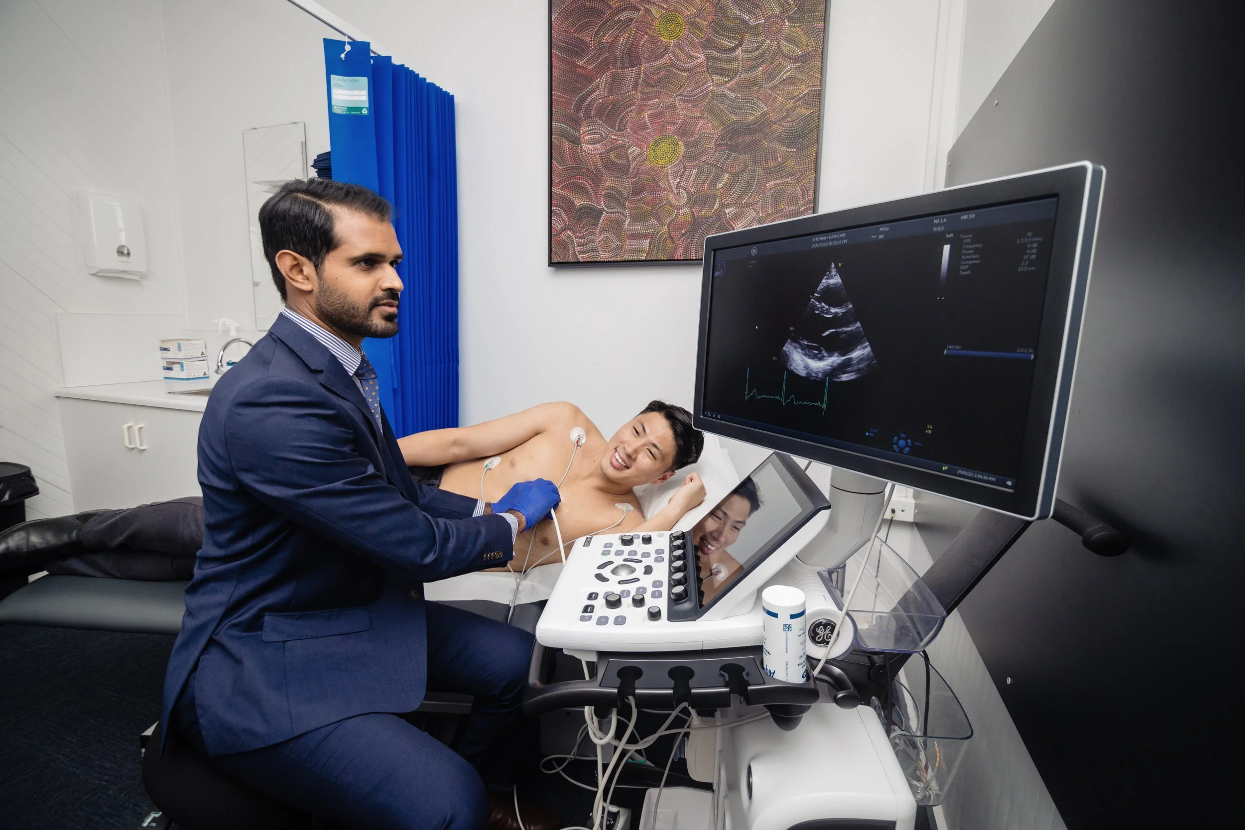
Our diagnostic tests are performed by experienced cardiac staff in a calm and professional environment. Most tests are non-invasive and designed to provide your cardiologist with important information about your heart health.